


Every year roughly 1,000 Canadians are diagnosed with amyotrophic lateral sclerosis (ALS, also known as Lou Gehrig’s disease). Some 4,000 Canadians are currently living with the disease.
There are few effective treatments available to slow the progression of ALS and, unfortunately, most sufferers die within two to five years of being diagnosed.
As neuroscience researchers, we are dedicated to finding ways to reduce the symptoms of ALS. In this article, we discuss the promising results of one of our recent studies.
ALS, a complex disease
ALS is a neurodegenerative disease. Sufferers gradually lose the ability to perform voluntary movements due to loss of communication between the nervous system and muscles.
Our scientific understanding of ALS has evolved considerably since the disease was first described by the neurologist Jean-Martin Charcot, in 1874.
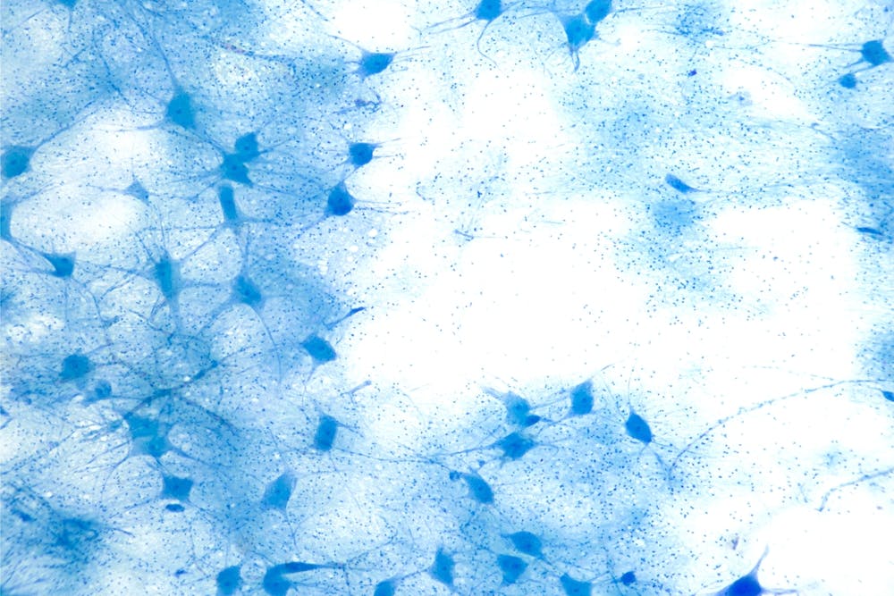
Today it is widely recognized that ALS affects several types of cells, including immune cells, muscle cells, glial cells (those that surround and insulate neurons) and neurons. However, for reasons that are still unknown, motor neurons, the cells responsible for controlling voluntary and involuntary movement, are particularly vulnerable.
Targeting motor neurons
Motor neurons have a complex morphology with projections that transmit information, resembling the branches of a tree. The neurons connect the brain and spinal cord to the muscles. These neuronal connections with the muscles are responsible for voluntary movements such as walking, speaking and eating.
In ALS, motor neurons degenerate and die, which leads to muscle weakness and, ultimately, paralysis. Because ALS is a complex disease with few effective therapies, there is an urgent need to better understand it in order to develop more therapeutic strategies.
Our research team in the Department of Psychiatry and Neuroscience at Université Laval is dedicated to understanding the early pathological changes that occur in motor neurons before they die. Our aim is to identify and develop therapeutic interventions that can reverse these early changes and slow the progression of the disease.
How do neurons lose their connectivity?
Using the mouse as a model, we carried out genetic modifications to recreate the motor and cognitive disability brought about by ALS.
In doing so, we discovered that changes in the morphology of motor neurons in the brain occur early in the disease process. This happens even before any changes in motor function have been seen.
We also discovered that a decline in motor functions was associated with the degeneration of motor neurons and the loss of synapses. Synapses are the places where motor neurons communicate, thus connecting the brain and spinal cord to the muscles.
Our study also found that astrocytes and microglia, the immune cells of the brain and spinal cord, were highly activated while synapse loss was underway and motor function was declining.
The over-activation of astrocytes and microglia, which can be described as neuroinflammation, changes the morphology of motor neurons and impairs their interconnectivity. These changes affect the neuronal network and disrupt the transmission of information.
Can we restore motor neuron connectivity?
We then wondered whether, by blocking neuroinflammation (for example, the activation of astrocytes and microglia), it would be possible to restore the morphology and connectivity of motor neurons.
In an attempt to answer this question, we tested the efficacy of a semisynthetic drug based on withaferin A, an extract of the ashwagandha plant which has been used for thousands of years in traditional Indian medicine.
The results of our study show that by blocking the activation of astrocytes and microglia, and thereby preventing inflammation, the morphology of motor neurons and their synaptic connections could be restored.
These promising results coincide with cognitive and motor improvements observed in our ALS mouse model.

Inflammation and loss of synapses are common to several neurodegenerative diseases, including ALS, Alzheimer’s and Parkinson’s disease.
As a result, therapeutic approaches that block inflammation and encourage the restoration of neuronal morphology and synaptic connections could have a wider application in the treatment of neurodegenerative diseases.
Future work by our team will aim to further develop treatment strategies that target inflammation and restore synaptic function in ALS sufferers.